Pain in your knees are troublesome and often influences your everyday life, like walking or climbing stairs. There are three different types of arthritis that can affect your knees. The most common type of arthritis is osteoarthritis (OA), a progressive condition where the cartilage between your bones, in this case the femur and tibia, degenerate over time. It is similar to getting grey hair, it is a completely normal part of aging. Osteoarthritis normally affects the “big” joints in the body that are subject to a lot of wear and tear, like the knees, hips and shoulders.
Rheumatoid arthritis is an inflammatory condition that affects not only one, but multiple joints, like the wrist and fingers. It is a systemic condition and disrupts the joint surfaces.
Post-traumatic arthritis develops after an injury directly to the knee. Post-traumatic arthritis doesn’t happen immediately after the trauma, it occurs mostly as an end result after years of repetitive damage to the structures inside the knee joint. This can happen because of a meniscus tear, ligament injury, fracture or surgery. Physiotherapists are qualified to distinguish between the different types of arthritis and help you to make the changes needed to live with knee joint osteoarthritis.
What is the knee joint made up of?

Your knee joint is a synovial hinge joint that connects the femur (thigh bone) to the tibia (shin bone). There are two joints that work together to bend and straighten the knee:
1) the tibiofemoral joint, between the femur and tibia, and the
2) patellofemoral joint, between the patella (kneecap) and the femur.
These two joints work together to form a hinge joint, that allows movement in only one plane, allowing the knee to bend and straighten. There is a very slight rotary component to knee movement with weight bearing too, but this is under unconscious control. This is important when the knee is loaded and moving, like during a single leg squat.
The bony ends have cartilage in between, similar to the discs in the spine. It acts as shock absorber and decreases friction when the bony ends move on top of one another. As time passes the cartilage can become thinner, almost like tyres that has done a lot of milage. This leads to less shock absorption and less “smooth” movement.
Stages of knee joint osteoarthritis: how bad is it?
Unfortunately osteoarthritis is a progressive disease and we cannot change what has already happened within the joint. We can however slow down the changes. This is what you can expect:
Stages of knee arthritis:
- Zero (0) is a normal, healthy knee.
- I is minor bony outgrowths or bony spurs
- II is mild, more bony spur growth but cartilage size remains normal. Healthy cartilage = healthy space between joints.
- III is moderate, cartilage damage and the space between joints begins to narrow. You will experience pain when you bend your knee.
- IV is severe. A lot of cartilage damage associated with a lot of pain and discomfort when you walk. If you are in this stage it also limits your every day activities, like driving, walking or climbing stairs.
Something interesting
The largest joint in our body, is the knee joint! It is quite vulnerable to injury as it has to bear a large amount of pressure while providing movement at the same time. When you walk, the load on your knees are equal to 1.5 times your body weight. While you climb stairs or run, it is equal to 3-4 times your body weight. When you squat, the load on your knees increases to about 8 times your body weight!

Causes of knee joint osteoarthritis
Osteoarthritis leads to cellular changes in all the joint tissues, including articular cartilage, subchondral bone, synovial membrane, ligaments and muscles. It is a degenerative disease that responds to mechanical stress, which leads to cartilage breakdown and bone remodeling, ultimately contributing to joint dysfunction. The main characteristic of increased inflammation is driven by a local cytokine reaction within the synovial fluid. The synovial fluid is a lubricant that allows smooth movement of the knee joint. This causes inflammation of the synovial membrane, damage to the cartilage and osteophyte formation.
Cytokines causes inflammation, usually in response to tissue injury. This makes tissues highly sensitive and stimulate nociceptors, that relay messages related to damage, as a sensation of pain via the spinal cord to the brain. These cells are present within the synovial fluid of someone suffering from osteoarthritis.
- Older age
- Gender – osteoarthritis is more common in women
- Previous knee joint injury – as far back as your school days
- Previous trauma (including surgery)
- Repeated stress on the joint – because of work or hobbies
- Genetics
- Bone or postural deformities
- Metabolic diseases – Diabetes or hemochromatosis (too much iron in your blood)
Osteoarthritis of the knee joint in stages
Stage 1
Some bone spurs will show on X-rays, this is pieces of bone that develop where the bones move on top of each other inside the joint. You will slight discomfort as a result of minor friction on the bones. A gradual increase in pain that develops slowly. The first thing many patients notice is pain and stiffness in the morning, that eases as you start getting active. The same thing happens when you are in one position for a long time, like at the movies or far drive, you feel stiff and sore this first few steps you take.
Treatment
Your doctor may prescribe analgesics or anti-inflammatory medications. A physiotherapist is able to recommend an exercise routine to help relieve your symptoms and slow the progression of your knee joint osteoarthritis.
Stage 2
X-rays will show bigger bone spurs, but the cartilage is healthy. The joint will be maintained. Synovial fluid is a lubricant present inside the joint that ensures smooth movement of the femur on the tibia. Bony spurs stimulate the synovial membrane to cause swelling.
Treatment
Treatment is aimed to relieve the pain. An exercise program, that includes low impact aerobics and strength training, will help support and protect your knee.
Stage 3
X-rays show decreased joint space between the tibia and femur. Damage to the cartilage, which protects the bones from scraping on each other, is present in patients with stage 3 knee joint osteoarthritis are likely to experience frequent pain when walking, running, bending and increased stiffness in the morning that takes longer to dissipate.
Treatment
Physiotherapy will aim to modify and change the load put on your knee joint, using joint mobilization and strapping techniques. Specific exercise prescription is necessary to prevent further damage.
Stage 4
X-rays show joint disruption and severe joint space narrowing. The cartilage is greatly affected and therefor the joint lubrication too. This makes the knee feel sticky and tight.
Treatment
An orthopaedic surgeon will make the decision to do a total knee replacement. Remember that replacing your arthritic knee won’t be the end of your osteoarthritis knee problems. It is just the start of the rehabilitation program.
Self tests for knee osteoarthritis
Diagnosis of knee joint osteoarthritis
Our therapists are skilled to take a thorough history and perform a clinical assessment, to make an accurate diagnosis. Osteoarthritis is a degenerative disease and management is key. We are able to lead you every step of the way.
Movement tests
Our physiotherapists will test the range of movement of your knee to identify what hurts. This enables us to know how much of the problem in caused by the joint surface and how much is caused by muscular restriction.
Contributing factors
We will also assess movement and stability of your hips and lower back, as imbalance from one area can lead to compensation strategies in another area.
Neurological assessment
If you have referred pins & needles or numbness we will also do a neurological assessment to identify which nerve is causing the symptoms. You can expect sensation, muscle power and a reflex hammer for these tests.
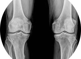
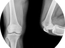
X rays
X rays are the image of choice to diagnose osteoarthritis because it shows the bones. We will be able to see any bone spurs, joint space, alignment and the quality of the bone. If you have older x rays, comparing the images is of great value to track disease progression. Increase in pain does not necessarily indicate a worsening of the disease process, it may be indicative of increase in joint dysfunction which can be managed with physiotherapy.
MRI
MRI is a costly image that can only be ordered by a specialist. It shows bone and soft tissue, giving information of what is going on inside the joint. Sclerosis, tissue thickening, or cysts, fluid filled pouch, will be visible with a MRI. It will not be necessary for diagnosing OA of the knee joint.
Why is my pain not going away?
Osteoarthritis is a progressive disease process. Nobody can change what has already happened within the joint. Physiotherapy is aimed at understanding how to manage your symptoms and slow the progression down, by increasing your muscle strength to decrease load on the joint itself. Doing too much will aggravate your symptoms and doing too little will increase joint stiffness and again cause pain. You have to find the balance between too much and too little in order to keep active, stay mobile and strong. We can help you find this balance.
It is normal to experience flare ups of pain. This is a short period of time where the symptoms increase and function may decrease as a result. This can be caused by over exertion, new movements or hobbies or even prolonged inactivity, like a long journey. All of this can increase inflammation within the joint. This increase in inflammation may feel warm and look red and swollen. This will limit movement and cause a “grainy” sensation on movement, almost like there is sand within the joint. The knee may make a noise, like clicking, cracking or popping sensation when moving. It may feel unstable when walking or climbing stairs.
Morning stiffness is very common with osteoarthritis. This is caused by the inflammation within the joint. Movement and activity improves the circulation to and within the joint and will ease the stiffness. It usually lasts approximately 30 minutes.
")
A big problem we see with knee joint pain
Being promised an easy fix. A lot of patients hopefully try a wide variety of treatment options, exercise regimes, medication, supplementations and weight loss strategies without understanding the pathology of osteoarthritis. Unfortunately there is not easy fix and no way to give you back the knees you had in your twenties. No supplement can reverse the damage. A good understanding of what is going on inside your knee joint, how to load it responsibly and what exercises to do is the best strategy to manage osteoarthritis of the knee joint.
Bandages or braces worn irresponsibly can actually cause weakness, because the muscles get lazy. A compression bandage applied to reduce swelling for a short duration is greatly beneficial. Wearing a brace constantly is not.
Chiropractic manipulation of a knee joint with osteoarthritis can be dangerous because of the physiological changes within the joint. Bony spurs and sclerotic ligaments may be damages with a forceful manipulation leading to even more instability of your knee.
Walking with a crutch or cane that is not measured for you specifically may cause more harm than good. It may affect your gait pattern to such an extend that you develop hip or back pain in the long run.
Physiotherapy treatment for knee joint pain
Unfortunately we cannot reverse the degeneration, it is not as simple as that. We can relieve a great deal of your pain and assist you in some lifestyle adaptations that will help you minimize your knee pain. In doing things differently you can slow down the degeneration process and limit painful episodes.
A good understanding of what is going on inside your knee joint, how to load it responsibly and what exercises to do when, is the best strategy to manage osteoarthritis of your knee joint. We can help you understand the pathology, how and when to do what, to manage your symptoms and slow down disease progression.
Treatment will include:
- electrotherapy, including laser and ultrasound, to help with pain relief
- myofascial release and dry needling to get rid of muscle spasm and tightness
- joint mobilisation to improve or restore the movement of the joints
- nerve mobilisations to restore the normal gliding movements of the nerves
- taping/strapping to support the area
- exercises to strengthen the stabilizing muscles around the knee
- advice and education to help you to understand your condition and know what you can change to be in control of your symptoms.
1st Phase: Protection & initial pain management
The first phase of your treatment will involve lots of information about your condition. We will explain the treatment plan and give you advice on everyday things to help manage your pain, from sitting positions to how to get up from a chair.
During this phase of treatment we will use joint mobilisations and myofascial release techniques to decrease your pain. Electrotherapy like ultrasound and laser will also help with pain relief at this stage.
We use strapping or taping to help support and protect the area while we start with gentle, pain free exercises. If you continue walking on the sore leg, you may start to limp. Even though limping eases the load from the knee, it influences the rest of the kinetic chain and you may start to experience back or hip pain too. The load placed on the knee can increase the inflammation during an acute flare up. It’s better to decrease the load on your knee, by avoid climbing stairs or long walks during this first phase. You could substitute your walking or jogging with swimming or cycling.
Ice helps with pain management and swelling, where heat helps with mobility in your knee. When you are experiencing pain and swelling it is good to use cold therapy (ice cubes wrapped in a towel around your knee joint) for about 10 minutes every two hours until the swelling subsides.
Heat improves mobility by easing tension in the muscles around the knee joint. You can use a hot water bottle or bean bag for 20 minutes 3 times a day. Be sure to put a towel between the heat pack and your skin.
2nd Phase: Establish pain free range of movement
During your examination and testing, it’ll become clear what you can do and what makes your pain worse. We identify factors that contribute to your pain, specific to your case.
The aim is to maintain the available movement in your knee joint. We use joint and neurodynamic mobilizations to maintain or gain range of movement. Pain management is still key during this phase and we will use Laser, Soft tissue mobilizations and Ultrasound therapies. Strengthening of the stabilizing muscles around your knee joint is also essential. Therefore, your physiotherapist will give you exercises to do at home to gain strength.
3nd Phase: Isometric exercises
If all movements of your knee is painful isometric exercises gives you all the benefits, without any of the pain.
An isometric exercise means that the muscles contract, but the joints don’t move. It may not feel like much, but the benefits are great. We use these muscle contractions to help with initial activation of the muscles around your knee.
4th phase: regain range of movement
Morning stiffness is a characteristic of osteoarthritis. During our initial assessment we find the movements that are limited and painful. If pain is what limits your movement we will use joint mobilisation, myofascial release and stretches to restore the movements of your knee.
If your degeneration is advanced or severe, there will be a loss of movement that isn’t reversible. Our treatment will focus on restoring the available range and then strengthening your muscles to maintain the range of movement we have gained . This muscle strength is very important for long-term improvements in your condition. The stability of your knee improves when the muscles are stronger, so your body doesn’t have to use muscle spasm and osteophytes to help with stability.
5th Phase: Concentric strengthening
After the initial phases of treatment you will have more movement of your knee. Now we start strengthening these ranges. A concentric contraction is when the muscle fibers shorten, allowing the joint to move. For example, the quadriceps muscle shortens when the knee is straightened.
As you progress through this phase and your muscles get stronger we will add resistance to the exercises, with weights or resistance bands. This ensures that the muscles will be strong enough to support your knee when you climb stairs or walk uphill.
6th Phase: Hip girdle strengthening
The hip girdle is made up of your pelvis, lower back and hip joints. Imbalance from the hip muscles can influence the health of your knees. During this phase of rehabilitation we will add exercises to strengthen your lower back and hips too. This approach ensures that the load be carried through the entire body to help the knees.
7th Phase: Testing for return to activity
Over the course of rehabilitation we will build up your strength and endurance to allow you to return to what you love doing. This is important to determine if your condition can withstand the repetitive loading without flare-ups. Our physiotherapist will guide you to reengage in safe increments and adjust your exercises where necessary. We’ll follow your progress and adapt where needed.
8h Phase: Sport Specific Training
This is the final stage of rehabilitation that can last anything from 2-4 weeks. Your physiotherapist will still continue with joint mobilization, myofascial release, trigger point release and electrotherapy modalities where needed, but functional rehabilitation is most important during this phase.
Depending on your sport, your physiotherapist will tailor specific exercises that will help strengthen the muscles pertaining to your sport. A successful outcome is when you have gained knowledge throughout the rehabilitation process and can participate at full power and speed.
Healing time for knee osteoarthritis
Osteoarthritis is a degenerative condition, so we cannot reverse or heal the damage that has all ready occurred. However, we can make a difference to your pain and quality of life. Our treatment plan and how often we need to see you, will depend on the severity of your condition and your current level of pain. We will need to see you to predict what amount of change can be expected from your unique case.
Usually if you have constant pain, we will need to see you twice a week, until your pain is under control. Thereafter we will make an appointment once a week to maintain the movement we have gained and to increase the intensity of your exercises. Then we may space our sessions to every second week or once a month. You may have flare ups in between sessions, this is no cause for concern, as we can treat as the need arises.
Without treatment or if you don’t finish your rehabilitation, the pain and stiffness will always return and get worse over time. At some point you will find that you have constant discomfort and or pain that isn’t relieved by rest. Working with your physiotherapist you can make sure that this doesn’t happen.
Another very important thing to remember is that even if your doctor has suggested surgery, you don’t have to wait until after your procedure to start physiotherapy treatment. Prehabilitation ensures that you are strong before surgery and can dramatically shorten your recovery after surgery.
Other medical treatments for knee arthritis
Blood tests can be ordered by your doctor to determine if there is a systemic cause for your knee joint pain. Inflammatory markers, C-reactive protein (CRP), is indicative of inflammatory processes in the body.
Your GP can prescribe medication to relieve your symptoms during an acute flare up. Cortisone or anti inflammatory medication will reduce the swelling. It is important to take medication that does not cause digestive side effects (constipation or burning), because you may be on the medication for an extended period of time.
Intra articular gel injections (Synvisc one) helps with joint lubrication and can last for up to six months.
No joint manipulation will take the cause of your symptoms away. Chiropractic manipulation of a knee joint with osteoarthritis can be dangerous because of the physiological changes within the joint. Bony spurs and sclerotic ligaments may be damages with a forceful manipulation.
No brace or knee guard will make your knee more stable. It may make your knee feel more stable, but would you not like to have faith in your own muscles to give you stability. The compression given by a knee brace can help reduce swelling, this tactile support will stimulate muscle activation in the short run. Regular, long term wear will have the opposite effect, and essentially make the muscle around your knee lazy and incapable of carrying the load.
Can excess weight influence my knee joint paint?
Losing weight, if you are overweight (BMI of 25 or higher), will definitely ease your knee joint pain. Interestingly enough not only because it lightens the load, but also because of the big role adipose tissue (fat cells) play in inflammation. The less adipose tissue you have, the less cells that can cause and sustain an inflammatory reaction. Less inflammation = less swelling = easier joint movement = less pain!
Surgery for knee joint osteoarthritis
Total knee replacement
The most common procedure offered for severe knee joint pain is a total knee replacement (TKR). Depending on your age, previous surgical interventions and your surgeon’s preference, a partial replacement may be considered (where only half of the joint is replaced). Even though you technically get a brand new knee, it still won’t be as good as the knee you had in your twenties, unfortunately. And even this is not a quick fix. It takes months of rehabilitation to make the surgery worth while. We can help you through this decision and with your rehabilitation. Please remember that the stronger you go into your surgery, the easier your recovery will be. So you can start “prehabilitaion” before you even have the date of your procedure.
It is a brutal procedure. You may opt for general anesthesia, but to prevent surgical complications, a lot of knee replacement are performed with an epidural. This means that you will be paralysed from the waist done and given a sedative for theatre, but you will be able to hear what is being done. The incision is made vertically down the front of your knee, the tell tale scar you may have seen before. Then the bottom end of the femur is removed and replaced with a stainless steel prosthesis, and the top end of the tibia is removed and replaced with a polyethylene surface for articulation. The knee will be tested in theatre to make sure it is aligned and mobile.
Post surgery rehabilitation
After the surgery the hard work starts. You will be helped out of bed the day after surgery and start with your rehabilitation. The hospital stay is usually one week, on until you can actively bend the knee to 90 degrees, completely straighten it and climb a stair. Once discharged from hospital you will follow up with your surgeon with certain intervals. Improving your range of motion and strength and endurance is of utmost importance now, otherwise it will all have been for nothing. Disciplined rehabilitation will get you the results you want. This can take up to 12 months after the surgery, depending on your goals. You may want to do another half marathon after all.
What else could the knee pain be?
Injury to the meniscus will have a traumatic incident and result in “locking” of the knee if there is meniscal displacement and an inability to straighten the leg.
Knee ligament injuries usually have a traumatic incident and instability because of it, expect a feeling of the knee giving way.
- Gout
Red, hot and swollen and mostly the result of some over indulgence, rich meaty stew and red wine.
Superficial pain will be focussed at the front of the knee and worse when bending.
- Knee bursitis
A fluid filled little balloon at the front of the knee after kneeling for a long time, scrubbing floors or gardening.
- Fracture
The patella (knee cap) can be fractured from a direct blow to the knee, severe pain, instability and a inability to move the joint after injury will follow.